
Don’t misdiagnose melanoma in diabetes - The new U.L.C.E.R acronym
- Ivan Bristow

- Jun 3
- 3 min read
The Problem of melanoma
Over the years I have written and published much about the issues of melanoma on the foot and its delayed diagnosis. Published studies highlight how melanoma on this part of the body is a problem for patients and practitioners alike. Delays in patient presentations arise for a diverse range of reasons including lack of visibility to the patient, not recognising serious disease [1], too busy to consult a doctor or fear over a diagnosis [2]. On the practitioner side, misdiagnosis is frequently an issue. Melanoma is documented to mimic many frequent, benign diagnoses [3, 4]. Clinically when there is a low index of suspicion, favours the more common conditions.
In the diabetic foot, potentially misdiagnosis issues are magnified with an increased risk of cutaneous complications such as ischaemia, infection and ulceration. From my own research, melanoma masquerading as diabetic foot ulcers is a common occurrence evident from the literature [5]. Consequently, myself, Michelle Reynolds (Postgraduate researcher at Health Sciences University) and Dr Matthew Young (Consultant Physician, Edinburgh Royal Infirmary) undertook a systematic review of the literature over 15 years to characterise the issue.
The Publication
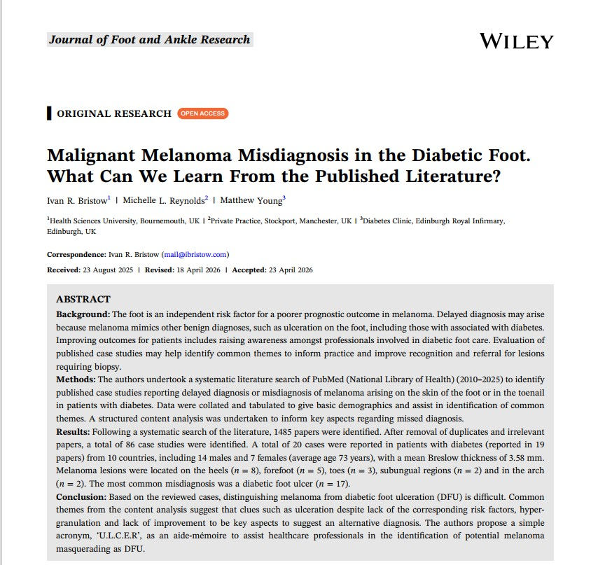
Published in the Journal of Foot and Ankle Research, we examined the characteristics of all case arising in the literature and compiled their characteristics. The vast majority of melanoma in the case series were initially diagnosed as diabetic foot ulcers. This was of concern as in many cases there were no identifiable risk factors for ulceration such as neuropathy or ischaemia.
We then went on the examine the reported pitfalls in each case. We undertook a content analysis of the discussion to identify themes around misdiagnosis and record the frequency of each. The analysis uncovered 5 main areas where misdiagnosis may occur. Consequently, from this data we have created and published a new acronym “U.L.C.E.R” as an aide-memoire below.
U |
Unusual – spontaneous bleeding or hypergranulation tissue, ulcer located in atypical area for ischaemic or neuropathic lesions.
|
L
|
Longevity – a lesion which is static or deteriorating despite treatment.
|
C
|
Colour – irregular colour within, or patchy pigmentation around the ulcer
|
E
|
Evolution or enlargement despite treatment
|
R |
Risk factors for diabetic ulceration maybe absent such as neuropathy, ischaemia or infection
|
Table 1: The U.L.C.E.R acronym [Bristow, Reynolds & Young 2026]
Personally, I notice there are many guidelines available for diagnosing and managing diabetic foot ulcers such as those from the National Institute for Health and Care Excellence and the International Working Group on the Diabetic Foot [6]. They discuss assessment – highlighting checking for vascular and neurological complications along with guidelines for management including antibiosis, off loading and dressing but fail to include details and considerations for non-healing lesions.
Biopsy is the only way to diagnose melanoma and other malignancies that may masquerade as ulcers. Inclusion of a sentence such as “If non-healing or deterioration, despite treatment, consider an alternative diagnosis and biopsy when the diagnosis is in doubt”, for example.
Ultimately, an acronym can be a useful aide-memoire and can simplify complex presentations:

The full free paper can be found on the Journal of Foot and Ankle Website here.
The Colour Infographic above can be downloaded by right clicking the image and selecting "save as".
References
1. Burke OM, Shivashankar V, Jaimes N, Kirsner RS: Acral Lentiginous Melanoma: Overcoming Diagnostic Delays Through Early Detection Strategies. J Am Acad Dermatol 2025, 94:567-575.
2. Xavier M, Drummond-Lage AP, Baeta C, Rocha L, Almeida AM, Wainstein AJA: Delay in cutaneous melanoma diagnosis: Sequence analyses from suspicion to diagnosis in 211 patients. Medicine (Baltimore) 2016, 95:e4396.
3. Soon SL, Solomon AR, Jr., Papadopoulos D, Murray DR, McAlpine B, Washington CV: Acral lentiginous melanoma mimicking benign disease: the Emory experience. J Am Acad Dermatol 2003, 48:183–188.
4. Bristow IR, de Berker DA, Acland KM, Turner RJ, Bowling J: Clinical guidelines for the recognition of melanoma of the foot and nail unit. J Foot Ankle Res 2010, 3.
5. Lyundup AV, Balyasin MV, Maksimova NV, Kovina MV, Krasheninnikov ME, Dyuzheva TG, Yakovenko SA, Appolonova SA, Schiöth HB, Klabukov ID: Misdiagnosis of diabetic foot ulcer in patients with undiagnosed skin malignancies. Int Wound J 2022, 19:871–887.
6. International Working Group on the Diabetic Foot: IWGDF Practical guidelines on the prevention and management of diabetic foot disease. Amsterdam: International Working Group on the Diabetic Foot; 2019.


