
Beyond the foot: considering extragenital lichen sclerosus in podiatry
- Michelle Reynolds
- Jul 31, 2025
- 10 min read
Introduction
How often do you get midway through a new patient consultation only to discover your patient has failed to initially disclose some vital medical history, as they didn’t feel it was relevant? Less frequently you may see a patient who you feel has overshared. With women’s health receiving more attention you may now encounter patients who are happy to divulge conditions affecting for example the anogenital area that you perhaps did not think were pertinent to podiatry consultations. However, they may be extremely important, and it is vital we start to think beyond the lower limb.
As podiatrists, we encounter a vast array of skin conditions affecting the lower limbs and feet. From fungal infections and common warts to calluses and pressure lesions, the typical presentations are often readily identifiable yet can be mis- or over diagnosed. The skin can be a canvas for numerous less common conditions, some of which may initially mimic more benign issues or appear in unusual ways. One such condition that warrants inclusion in every podiatrist's differential diagnosis is Lichen Sclerosus (LS). While primarily known for its impact on the anogenital area, LS can manifest extragenitally, and crucially for podiatrists, this extragenital form (ELS) can affect the feet [1-2] .
Recognising ELS on the foot is not only important for appropriately managing the local symptoms but also because it can potentially herald the onset, or co-exist with more severe anogenital disease, which carries a risk of malignancy [3-4] .
What is Lichen Sclerosus
LS is defined as an uncommon, chronic, lymphocyte-mediated, inflammatory dermatosis [1], yet it is estimated to affect around 1 in a 100 women, and may be more common due to the low rate of recognition and delay in diagnosis. The most frequent clinical variant of LS presents with porcelain white, pruritic, and atrophic plaques on the genitalia [1, 4].
LS most commonly affects women around the time of the menopause, but can occur, although less frequently, in men and children. It is a chronic inflammatory condition which can cause intense itching and dyspareunia and can have a dramatic effect on health quality of life, with particular respect to intimate relationships.
Diagnosis is commonly delayed [5], often by years, leading to worse long-term outcomes, such as neoplasm formation, surrounding cutaneous or mucosal changes and disfigurement and urogenital issues. Misdiagnoses frequently occur with LS initially being confused with thrush, eczema and other inflammatory skin conditions. Fissures, ulcerations and resultant infections can make everyday life difficult and uncomfortable.
What causes Lichen Sclerosus?
Autoimmune, infectious and genetic aetiologies have been suggested but ultimately the underlying cause is unknown. There is no known cure. Long term management is primarily with potent topical steroids. Without that ongoing treatment, LS can progress and start to cause structural changes to the vulva, including atrophy, labial and clitoral fusion, and scarring which are often irreversible.
Lichen Sclerosus can affect the skin elsewhere
However, LS is not strictly confined to the anogenital region. Extragenital LS (ELS) occurs in a minority of patients [1, 6]. The proportion of patients with ELS varies in reports, noted as approximately 15% to 20% of individuals with LS [1,4,6]. ELS can occur simultaneously with the genital form [4,7]. Interestingly, in some cases, only the extragenital form is observed, described as occurring in isolation in approximately 6% of patients [4, 7]. Common extragenital sites include the neck, shoulders, and upper portion of the trunk [4, 7, 8]. ELS cutaneous manifestations are rare and can vary widely in presentation [9].

Extragenital Lichen Sclerosus on the Foot
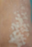
ELS starts with asymptomatic to mildly itchy white papules. These papules then merge into well demarcated erythematous plaques and become atrophic, appearing as ivory white patches with a tendency to scar [4]. It resembles being splashed with white wash. Early lesion may have comedone like plugs. Keratotic warty like lesions, sclerotic plaques, purpura, fissuring, milia, telangiectasia and bullae have all been reported.
While ELS can occur on various parts of the body, involvement of the foot is rare [1, 2, 3, 10, 11]. Several reports detail presentations of LS on the lower limbs and feet (dorsal and plantar aspects), illustrating the diverse ways this condition can appear in podiatric practice.
Symptoms may be absent with patients presenting due to the appearance alone, but lesions can cause intense pruritus, pain on weightbearing or palpation, and/or burning.
Lesions on the foot have been reported as presenting with:
Erythematous atrophic plaques and bullae [1]
Multiple white hyperkeratotic papules and scaling [3, 12]
Asymptomatic punctate hypopigmented atrophic macules [6]
Hypopigmented atrophic plaques with follicular plugging (which can occur along the lines of blaschko) [8]
Erythematous well demarcated sclerotic plaques with pale areas and yellowish translucent plaques [10]
Folliculocentric shiny atrophic papules coalescing into reticulated plaques [11]
White lichenoid plaques on the dorsum of the foot [13]
White-yellow papules and plaques, comedo-like openings and cribriform appearance [14]
One case study reported a 15 year history of chronic plantar ulceration with severe nail dystrophy resulting in the complete absence of a nail plate [15]

The Diagnostic Challenge: Mimicking Common Foot Conditions
The varied clinical presentations of ELS on the feet, particularly in its early stages can make it difficult to diagnose [9, 14]. The lesions might resemble other dermatoses commonly seen on the feet. According to the literature, in its early stages, extragenital LS may resemble morphoea, lichen planus, chronic eczema, vitiligo, and flat warts [14].
Consider the potential for confusion with:
Warts: Keratotic papules or warty-like plaques could easily be mistaken for viral warts
Eczema/Dermatitis: Scaling, pruritus, and erythema are common features of various forms of dermatitis affecting the feet
Calluses/Corns: Hyperkeratotic papules might be dismissed as simple pressure-related lesions
Fungal Infections: Scaling and altered skin texture could be misinterpreted as tinea pedis
Vitiligo: Hypopigmented macules could potentially be confused with vitiligo, although LS typically involves atrophy and changes in texture not seen in vitiligo. (Note: Vitiligo and LS are both autoimmune conditions and can be associated)
Stucco keratosis
This potential for misdiagnosis underscores the importance of maintaining a high index of suspicion when encountering unusual or treatment-resistant lesions on the feet [16]. Lesions of the hands and feet, located at sites that may also be susceptible to recurrent friction and trauma, could potentially develop due to the Koebner phenomenon, where new lesions appear at sites of injury. This has been described in ELS [3-4]. Recognising this possibility further highlights the need for careful examination and diagnosis.
Dermoscopy
While clinical examination is the starting point, dermoscopy has emerged as a valuable diagnostic tool for various skin conditions, including ELS. Dermoscopy of ELS lesions can offer crucial insights by revealing characteristic findings [14].
Early lesions may display the following dermoscopic features: white structureless areas, comedo-like openings, keratotic plugs and scale. Lesions may display the following dermoscopic features: white structureless areas, which are suggestive of the sclerosed dermis [17], comedo like openings, keratotic plugs, scale, linear or dotted vessels, pseudo pigment like pattern [6, 14]. More advanced lesions may show chrysalis structures, bright white patches, dotted, commalike or hairpin vessels [4, 9, 14].
Histopathology
Despite the utility of clinical examination and dermoscopy, the definitive diagnosis of LS, including ELS on the feet, typically requires histopathological examination [4] [11] [16]. A skin biopsy should be performed to confirm the diagnosis [11] [16].
Histopathology of LS characteristically shows thinning of the epidermis, basal cell degeneration, a band-like lymphocytic infiltrate in the upper dermis, and replacement of the normal papillary dermis with homogenized collagen or sclerosis [1, 3, 8, 11, 12, 14]. However, as mentioned earlier, in early forms, the hallmark features of sclerosis and atrophy may be absent, making the histopathological diagnosis, like the clinical diagnosis, potentially challenging [14, 18]. This further reinforces the need for a high index of suspicion and correlation of clinical, dermoscopic, and histopathological findings [11, 16].
The Critical Link: ELS and Genital Disease
Beyond the local symptoms and diagnostic complexity on the foot, there is a significant reason why podiatrists must be aware of ELS: its potential association with anogenital LS. While many cases of ELS, particularly on acral sites, may remain isolated to those areas [3], there is evidence suggesting that extragenital involvement can sometimes precede the development of genital disease [3].
This observation is crucial. If a podiatrist identifies ELS on a patient's foot, it provides an opportunity to prompt urgent referral for further examination of the anogenital area. This early detection of genital LS can be immensely important for the patient's long-term health and well-being [3].
Genital LS, which is the most common presentation [9], can be a significantly debilitating condition [5] . It causes characteristic porcelain-white plaques that can be associated with atrophy, erosion, purpura, and fissuring of the vulva and perianal skin [19]. For affected individuals, severe, intractable pruritus is a very common and distressing symptom [19]. The scarring and architectural changes caused by genital LS can lead to significant functional impairment and impact quality of life.
The Malignancy Risk: A Compelling Reason for Vigilance
One of the most serious implications of genital LS is its association with an increased risk of developing vulval squamous cell carcinoma (SCC) [3-4] . While the risk of malignant transformation in ELS is generally considered to be negligible, case reports with possible malignant transformation have been described in non-genital sites [4]. However, one source states that squamous cell carcinoma arising in extragenital lichen sclerosus is rare and has not been reported to involve lesions of lichen sclerosus in acral sites to date [3-4].
The key takeaway for podiatrists here is not necessarily the malignancy risk of lower limb lesions, but the opportunity that identifying an ELS lesion on the foot provides to screen for the presence of genital LS. Since genital LS carries a definite and serious risk of squamous cell carcinoma, diagnosing ELS on the foot allows for earlier detection and management of potentially more severe genital disease. This early intervention in genital LS can potentially reduce the patient’s risk of developing scarring and squamous cell carcinoma of the genitalia. Therefore, recognising ELS on the foot is not just about managing a skin condition on the extremity; it's about contributing to the holistic care and potentially preventing life-altering complications associated with genital disease.
Management and the Importance of Referral
For a podiatrist, identifying ELS on the foot means more than just initiating topical treatment for the local lesion. It necessitates a broader approach. Considering the potential link to genital disease and the associated malignancy risk, patients with suspected or confirmed ELS on the foot should be referred for a comprehensive dermatological or gynaecological evaluation to assess for and manage any concurrent or developing genital involvement [3].
Furthermore, LS is increasingly understood to be an autoimmune condition with a genetic predisposition. It has been associated with several other autoimmune diseases. These associations include autoimmune thyroid disease (such as Hashimoto's thyroiditis), pernicious anaemia, alopecia areata, psoriasis, and vitiligo [19]. Screening for thyroid disease and pernicious anaemia is warranted in patients with vulvar LS [19]. While these guidelines are specifically for vulvar LS, the autoimmune link suggests that patients with ELS, including acral forms, might also benefit from evaluation for associated autoimmune conditions. This highlights the importance of a multidisciplinary approach and referral for appropriate screening and management of co-existing conditions.
Conclusion: Be Alert for the Unexpected
In summary, while LS most commonly affects the anogenital region, it is a chronic inflammatory dermatosis that can manifest extragenitally. ELS on the foot is a rare entity, but its presentation can be remarkably varied. These diverse appearances, particularly in early stages before overt sclerosis and atrophy are present, can mimic numerous common foot conditions, making diagnosis challenging for podiatrists.
Crucially, the identification of ELS on the foot is important because it can, in some cases, precede the development of anogenital LS. Genital LS is a debilitating condition causing significant symptoms. More significantly, it carries a notable risk of malignant transformation into squamous cell carcinoma. While SCC arising in ELS on the foot appears to be exceedingly rare, diagnosing ELS at this site provides a vital opportunity to screen for and manage the higher-risk genital disease, potentially reducing the patient's risk of future scarring and malignancy in that area.
Therefore, it is imperative that podiatrists are aware of Lichen Sclerosus, understand that it can affect the feet, and include extragenital LS in their differential diagnosis when evaluating unusual, atypical, or treatment-resistant skin lesions on the lower limbs and feet. A high index of suspicion, especially for presentations that don't fit the typical picture of common foot dermatoses, coupled with consideration for dermoscopy and timely referral for biopsy and dermatological evaluation, can lead to earlier diagnosis and better outcomes for patients. By being vigilant podiatrists can play a crucial role in identifying a condition with broader implications for patient health.
Author Information:
Michelle Reynolds MSc (Derm) MRCPod is a podiatrist in private practice, Marple, Stockport.
For more information about Lichen Sclerosus, please use the links below:
References
1. Herz-Ruelas, M.E., et al., Acral bullous lichen sclerosus intolerant to UVA-1 successfully treated with narrowband ultraviolet B phototherapy. Photodermatol Photoimmunol Photomed, 2019. 35(5): p. 378–380.
2. Steff, M., et al., [Acral lichen sclerosus et atrophicus]. Ann Dermatol Venereol, 2008. 135(3): p. 201–4.
3. Heibel, H.D., A.R. Styles, and C.J. Cockerell, A case of acral lichen sclerosus et atrophicus. JAAD Case Rep, 2021. 8: p. 26–27.
4. Arif, T., R. Fatima, and M. Sami, Extragenital lichen sclerosus: A comprehensive review. Australas J Dermatol, 2022. 63(4): p. 452–462.
5. Conte, S., et al., Clinical presentations and complications of lichen sclerosus: A systematic review. JDDG: Journal der Deutschen Dermatologischen Gesellschaft, 2025.
6. Lee, S.B., et al., Acrosyringeal variant of extragenital lichen sclerosus et atrophicus. J Cutan Pathol, 2020. 47(11): p. 1039–1041.
7. Arif, T., Blaschkoid extragenital lichen sclerosus: an exceedingly rare presentation. Dermatology Review/Przegląd Dermatologiczny, 2024. 111(1): p. 62–65.
8. Diwan, N.G. and P.A. Nair, Extragenital lichen sclerosus et atrophicus along the lines of Blaschko. Indian Dermatology Online Journal, 2015. 6(5): p. 342–344.
9. Burshtein, A., J. Burshtein, and S. Rekhtman, Extragenital lichen sclerosus: a comprehensive review of clinical features and treatment. Arch Dermatol Res, 2023. 315(3): p. 339–346.
10. Vink, L. and T.M. Starink, Bullous acral lichen sclerosus with milia. Clin Exp Dermatol, 2014. 39(3): p. 400–1.
11. Al-Husain, K.M., et al., Acral Folliculocentric Extragenital Lichen Sclerosus: A Case Report. Clin Cosmet Investig Dermatol, 2024. 17: p. 1633–1636.
12. Viana Fde, O., et al., Acral lichen sclerosus et atrophicus--case report. An Bras Dermatol, 2011. 86(4 Suppl 1): p. S82–4.
13. Iraj, E. and E. Ali, Koebnerization in a Woman with Extragenital Lichen Sclerosus. Iranian Journal of Dermatology, 2008. 11(2): p. 86–88.
14. Alvarez-Rubio, F.J. and V.M. Tarango-Martinez, Acral Extragenital Lichen Sclerosus and Its Dermoscopic Findings. Cureus, 2024. 16(10): p. e70995.
15. Gualdi, G., et al., Nested Graft for Acral Lichen Sclerosus of the Feet: A Surgical Treatment for an Inflammatory Disease. Plastic and Reconstructive Surgery – Global Open, 2016. 4(3): p. e633.
16. Magana, M. and C.E. Marquez-Maldonado, An unusual presentation of extragenital lichen sclerosus-An extensive keratotic variant. Skin Health Dis, 2024. 4(3): p. e341.
17. Bhatia, D., D. Aggarwal, and A. Budania, Extragenital Lichen Sclerosus - Morphea Overlap within the Same Lesion: A Rare Occurrence. Indian Dermatology Online Journal, 9900: p. 10.4103/idoj.idoj_543_24.
18. Baklouti, M., et al., Extragenital Lichen Sclerosus: A Retrospective Study of 17 Patients. Indian J Dermatol, 2022. 67(2): p. 146–149.
19. Guttentag, A., et al., A Guide to Screening for Autoimmune Diseases in Patients With Vulvar Lichen Sclerosus. Australasian Journal of Dermatology, 2025.